SD-TDA combines the sensitivity and resolution of electron microscopy (EM) with the simplicity and speed of DLS, offering unmatched accuracy at these dimensions.
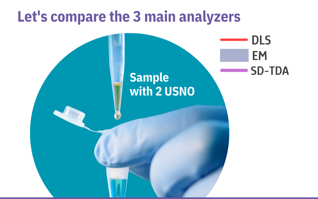
Discover the power of SD-TDA below two typical comparative examples.
Example 1
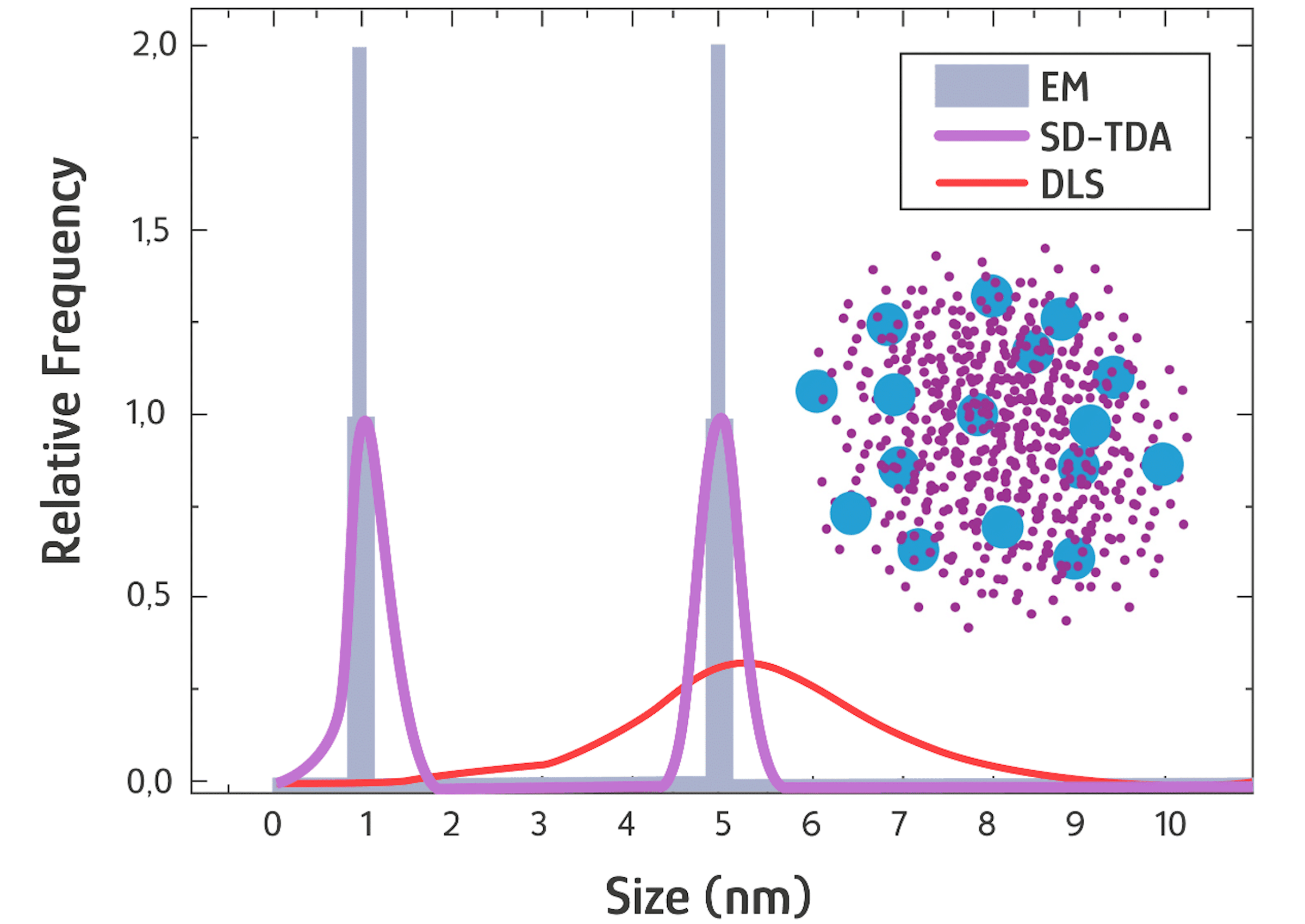
Mixture of 2 populations with a particle size ≤ 5 nm of equal volume fraction (polymer, proteins, USNP*, amino acids, peptides, core nanoparticles, etc.)
Open example details 1
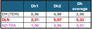
The Dh and PSD are substantially the same between EM and SD-TDA for the two measured species (approximately 1 nm and 5 nm). Both methods show size distribution histograms with two well-separated peaks and a return to baseline. DLS, however, cannot resolve these two species into distinct peaks due to its limited resolution for such small sizes (possibly a limitation of the measuring principle).
While the size measurement of the 5 nm species is accurate, the1 nm species is significantly overestimated at 2.3 nm.
The size distribution is very broad, resulting in an average Dh measurement of 5.3 nm, compared to an estimated Dh of 3 nm (perhaps due to traces of dust in the sample?).
Example 2

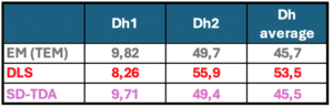
Mixture (90% v/v) of vesicles (LNP, liposomes, EVs, etc.) of 50 nm size in the presence of 10% of USNO* of 10 nm size (active ingredients, micelles, biomolecules, etc.)
Open example details 2
The Dh values are similar in EM and SD-TDA for the two species (10 nm and 50 nm), with high resolution comparable to that observed for smaller nanoparticles in the first example. In DLS, the smaller species at 10 nm are detected but with lower precision (8.26 nm instead of 10 nm). The size distribution in DLS remains broader for each species compared to EM and SD-TDA.
Additionally, the measured size for the 50 nm species shifts towards a higher Dh (55.9 nm instead of 50 nm), resulting in an overestimated average Dh of 53.5 nm, compared to 45.7 nm in EM. Note that the sharp Gaussian curve typically observed in DLS disappears when the data is converted to linear units, as shown in this example.
In the ever-evolving field of nanomaterial characterization, Size Dispersion – Taylor Dispersion Analysis (SD-TDA) has emerged as a groundbreaking technique.
If you are a fan of Electron Microscopy (EM, such as TEM, SEM, Cryo-TEM) or Dynamic Light Scattering (DLS), it’s time to explore why SD-TDA could surpass these methods in terms of precision and practicality.
Edge Over Electron Microscopy:
SD-TDA achieves a resolution comparable to electron microscopy in native and liquid media, eliminating the need for sample preparation and drying. It efficiently analyzes a large number of particles from a small volume, providing superior statistical value. The rapid measurement process (under a few minutes) requires minimal expertise for data interpretation.
Edge Over DLS:
SD-TDA offers significantly higher sensitivity and resolution than DLS when measuring the size of very small nano-objects, even in the presence of contaminants such as dust. Each particle’s contribution to signal intensity ensures uniform sensitivity regardless of size, unlike DLS, which detects larger particles more effectively than smaller ones in a mixture. This capability allows for reliable concentration measurement of each species by size. When paired with the Taylorsizer, which can autonomously process up to 50 samples, this technique becomes an excellent tool for high-throughput screening (HTS).
What About Other Measurement Techniques?
SD-TDA enables much faster measurements than analytical ultracentrifugation (AUC), A4F-MALS, or SEC-MALS, particularly for sizes below 50 nm, where sedimentation and retention times are typically long, expensive, and not suitable for HTS. Additionally, measurement techniques using light scattering, such as MALS or Nanoparticle Tracking Analysis (NTA), suffer from a lack of sensitivity for very small nano-objects.
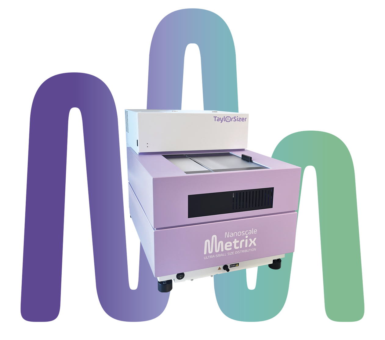
TaylorSizer (TM)
The ultimate UltraSmall size distribution analyzer
Are you also struggling to measure nano-objects from 0.1 nm to 50 nm in polydisperse mixtures?
Our team will enjoy to perform a demonstration and answer your questions.
We will respond as quickly as possible